

A Single Additional Dark-Field Image Reveals Hidden Pathological Details
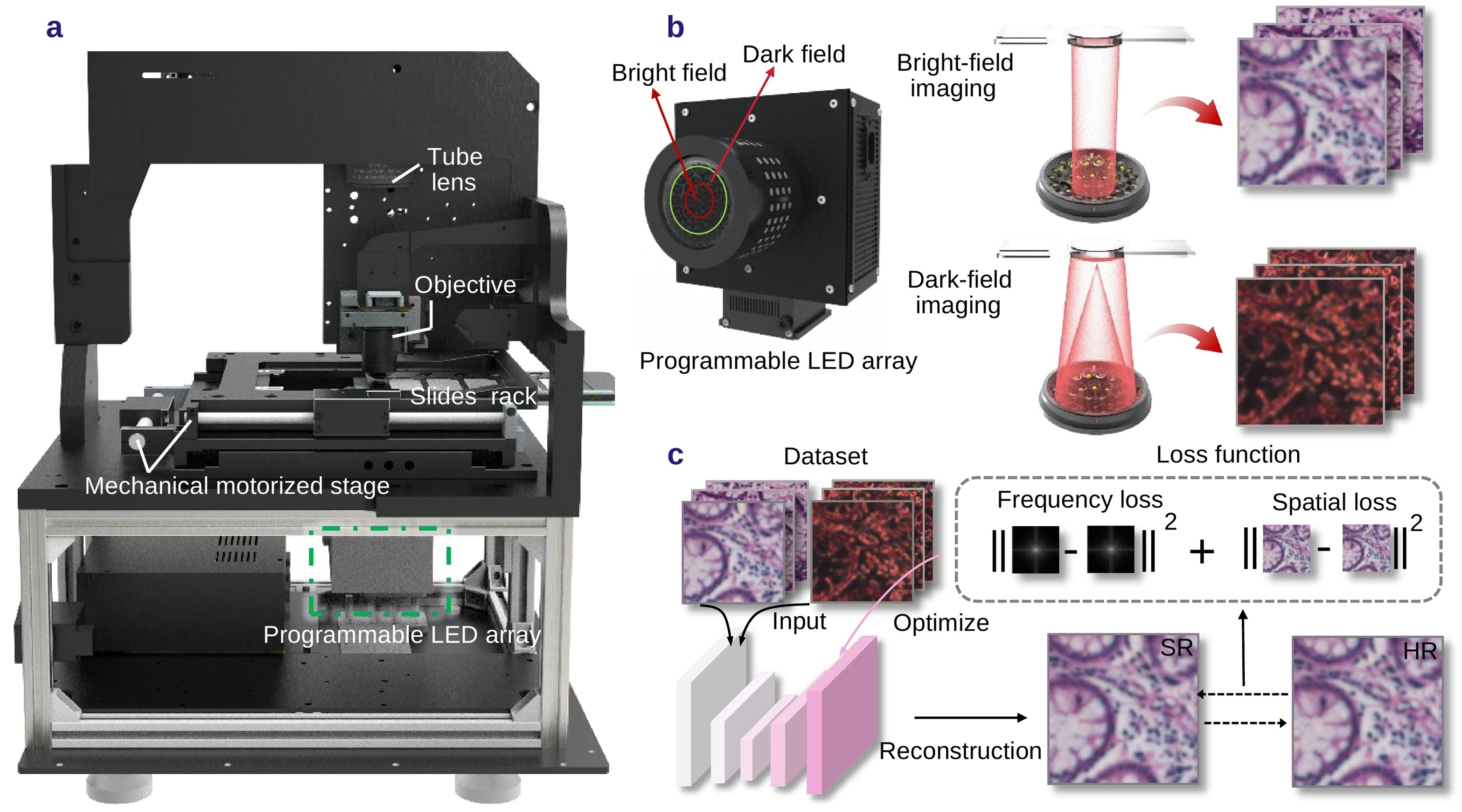
A programmable LED illumination source rapidly switches between bright-field and dark-field modes to acquire multimodal images. By integrating complementary information through a physics-guided deep neural network, the framework enables high-fidelity supe
Hybrid bright-dark field computational imaging enhances digital pathology resolution
CHENGDU, SICHUAN, CHINA, September 8, 2026 /EINPresswire.com/ -- Digital pathology relies on high-resolution imaging for accurate cancer diagnosis, yet high-magnification systems remain slow and costly for large-scale screening. Meanwhile, standard AI super-resolution often generates hallucinated cellular artifacts that risk diagnostic accuracy. To overcome this, researchers developed a hybrid bright-dark field computational framework using fast LED illumination to provide physical guidance. This physics-guided approach doubles spatial resolution, reduces reconstruction artifacts by 84%, and boosts AI cancer screening sensitivity by 11.14%.With the rapid development of medical imaging technologies and artificial intelligence, digital pathology has gradually become an important tool for advancing modern precision medicine. Whole-slide imaging (WSI) technology digitizes entire tissue sections through high-resolution scanning, transforming traditional microscopic observation into digital images that can be stored, analyzed, and shared. It provides a critical data foundation for disease diagnosis, tumor detection, and intelligent pathological analysis.
In recent years, with the rapid advancement of artificial intelligence algorithms in medical imaging, the importance of high-quality digital pathology images for improving computer-aided diagnosis has become increasingly evident. However, high-resolution imaging typically relies on expensive high-numerical-aperture (NA) optical systems, which suffer from slow scanning speeds and high hardware costs. The trade-off between resolution and efficiency has become a key bottleneck limiting the further development of digital pathology, particularly for large-scale pathological screening.
This bottleneck has been overcome by image super-resolution (SR) technology, which offers a promising solution by computationally recovering high-resolution information from low-resolution images. In recent years, deep learning-based single-image SR methods have achieved rapid progress. However, the limited information contained in a single low-resolution image often leads to fundamental challenges in reconstruction: regression-based approaches tend to produce over-smoothed results with missing fine textures, while generative models can improve visual quality but may introduce hallucinated artifacts. In pathological imaging, such artificial cellular structures may directly affect diagnostic reliability, severely limiting the clinical translation of conventional SR techniques.
Researchers have further developed reference-based SR (RefSR) methods to address the insufficient information problem of single-image SR. By introducing additional image information, these approaches provide auxiliary constraints for low-resolution image reconstruction and have demonstrated promising performance in medical imaging and live-cell imaging applications. Through integrating information from different imaging modalities, RefSR methods can exploit multi-dimensional observations to improve structural recovery. However, RefSR methods remain largely unexplored in digital pathology, and a key challenge lies in obtaining physically meaningful information strongly correlated with pathological structures while minimizing hardware modifications, thereby providing reliable prior constraints for SR reconstruction.
To address the instability of conventional single-image SR methods caused by the lack of effective physical constraints, a research team led by Professor Qian Chen and Professor Chao Zuo at Nanjing University of Science and Technology, in collaboration with Professor Juergen W. Czarske from TU Dresden, developed a novel digital pathology resolution enhancement method that integrates hybrid bright-dark field imaging with physics-guided deep learning, termed the Hybrid Bright-Dark Field Resolution Enhancement (HBDF-RE) framework. This work is made available online on August 27, 2026, in the Early View section of Opto-Electronic Advances.
The method employs a programmable LED illumination system to rapidly switch between bright-field and dark-field modes at each scanning position, acquiring a paired bright-dark field image within approximately 1/15 second. "The dark-field image provides scattering and edge-sensitive contrast as physical guidance, enabling the deep neural network to reconstruct high-resolution images from low-NA bright-field observations with performance approaching high-NA imaging," explained Prof. Chen.
By incorporating multimodal feature fusion, spatial attention mechanisms, and spatial-frequency joint constraints, HBDF-RE achieves high-fidelity reconstruction of cellular structures while effectively suppressing over-smoothing and artificial textures. With only one paired bright-dark field acquisition, the proposed method achieves approximately 2.1× spatial resolution enhancement, producing high-quality digital pathology images comparable to high-NA imaging without complex hardware upgrades, while improving computational efficiency by approximately 11%.
In the experiments, the researchers first applied HBDF-RE to an AI-assisted cervical cancer screening task. The results showed that the diagnostic sensitivity of the AI-assisted cervical cancer screening model based on HBDF-RE-reconstructed images increased by 11.14% compared with the original low-resolution images, with particularly significant improvements observed in clinically ambiguous lesion categories. Furthermore, in downstream analysis tasks such as gland segmentation, HBDF-RE reconstruction consistently outperformed existing pathology resolution enhancement methods, demonstrating its potential to improve intelligent pathology analysis and AI-assisted clinical diagnosis.
Furthermore, the researchers performed large-field SR reconstruction on human thymus tissue WSIs. Although the original low-NA scanned images enabled rapid coverage of large tissue areas, some nuclear boundaries and fine pathological structures remained unclear due to limited resolution. By leveraging the complementary information provided by bright-field and dark-field imaging, HBDF-RE successfully recovered fine tissue structures without requiring a high-magnification objective, achieving substantial improvement in spatial resolution capability. Compared with representative single-image SR methods, HBDF-RE further improved the peak signal-to-noise ratio by approximately 3.2 dB and reduced reconstruction artifacts by approximately 84%, demonstrating strong potential for large-scale digital pathology image enhancement.
By integrating physics-informed constraints with deep learning-based computational reconstruction, HBDF-RE leverages the complementary information provided by bright-field and dark-field imaging to effectively enhance low-NA pathology images without requiring costly optical upgrades. This approach overcomes the long-standing trade-off between resolution and scanning efficiency in conventional digital pathology systems.
"While maintaining the advantages of rapid large-field imaging, HBDF-RE requires only one additional dark-field acquisition at each scanning position, introducing minimal system complexity while achieving cellular structural representation comparable to high-NA imaging systems," remarked Prof. Chen.
In the future, combined with automated WSI platforms and real-time deep learning inference acceleration, this method has the potential to integrate high-fidelity resolution enhancement into routine pathology workflows, providing an efficient and reliable imaging tool for large-scale cancer screening, precision pathological diagnosis, and AI-assisted clinical decision-making.
***
Reference
Title of original paper: Learning from Hybrid Bright-Dark Field Imaging for Resolution-Enhanced Digital Pathology
Journal: Opto-Electronic Advances
DOI: 10.29026/oea.2026.260060
About Opto-Electronic Advances
Launched in March 2018, Opto-Electronic Advances (OEA) is an open-access, peer-reviewed SCI journal. Indexed in major databases including SCI, EI, and Scopus, it is guided by an international editorial board spanning 17 countries. OEA provides a fast-track platform for high-impact empirical and theoretical research across optics, photonics, and optoelectronics. Key focus areas include light sources, sensors, nanophotonics, plasmonics, biophotonics, optical imaging, intelligent optics, and optical communications, catering to researchers, academicians, and industry professionals seeking cutting-edge developments in light-driven technologies.
Website: https://www.oejournal.org/oea/en/
About Smart Computational Imaging Laboratory at Nanjing University of Science and Technology
The Smart Computational Imaging Laboratory (SCILab), at Nanjing University of Science and Technology (NJUST) is affiliated with the National Key Discipline of Optical Engineering. The laboratory is supported by the Ministry of Education’s Changjiang Scholars Innovation Team “Spectral Imaging and Information Processing” led by Prof. Qian Chen and the first batch of National Huang Danian Teacher Teams in Chinese universities. Prof. Chao Zuo, the academic leader of SCILab, is a Distinguished Professor of the Ministry of Education’s Changjiang Scholars Program and a Fellow of SPIE, Optica, and the Institute of Physics (IOP). Relying on several high-level research platforms, including the National Key Laboratory of Extreme Environment Optoelectronic Dynamic Measurement Technology and Instruments, SCILab has long been dedicated to fundamental theories, key technologies, and engineering applications of intelligent computational imaging. Its research covers optical imaging mechanisms, computational reconstruction, precision measurement, and artificial intelligence-enabled imaging systems, with broad applications in life sciences, biomedical diagnosis, and intelligent manufacturing. In recent years, the team has published more than 300 SCI-indexed papers, including over 50 cover papers in leading international journals such as Advanced Optical Materials (AOE), Light: Science & Applications, Optica, Advanced Photonics, and PhotoniX, as well as more than 30 ESI highly cited or hot papers. The team’s achievements have been widely reported by major media outlets, including CCTV’s Focus Report, People’s Daily, and Optics & Photonics News.
Website: www.scilaboratory.com
Funding information
This work was supported by the National Key Research and Development Program of China (2024YFE0101300, 2022YFA1205002, 2024YFF0505603, 2024YFF0505600), National Natural Science Foundation of China (62227818, 62361136588, 62305162, U21B2033, 62505136, 62575139), Fundamental Research Funds for the Central Universities (30923010206), Fundamental Research Funds for the Central Universities (2024202002), National Key Laboratory of Plasma Physics (JCKYS2024212804), and Open Research Fund of Jiangsu Key Laboratory of Spectral Imaging & Intelligent Sense (JSGPCXZNGZ202401).
Siyi Ma
Institute of Optics and Electronics, CAS
oej_publishing@ioe.ac.cn
+86 19136186296
Legal Disclaimer:
EIN Presswire provides this news content "as is" without warranty of any kind. We do not accept any responsibility or liability for the accuracy, content, images, videos, licenses, completeness, legality, or reliability of the information contained in this article. If you have any complaints or copyright issues related to this article, kindly contact the author above.
